EEG Spike Detection: What Clinicians Need to Know
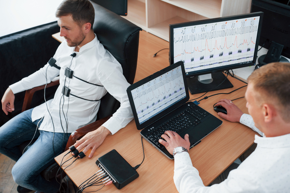
If you’ve spent any significant time reading EEG recordings, you already know the challenge. Hours of signal data, subtle waveform variations, and the constant cognitive load of distinguishing true epileptiform activity from artifacts, normal variants, and noise. Manual review is the gold standard — and it’s also exhausting, time-intensive, and inherently inconsistent across reviewers and sessions.
EEG spike detection sits at the intersection of clinical neurology, neurophysiology research, and increasingly sophisticated computational methods. Whether you’re a clinical neurophysiologist managing a high-volume epilepsy monitoring unit, a researcher building datasets for seizure prediction models, or a neuroscience graduate student trying to make sense of your first EEG dataset — this blog is written for you.
The goal here isn’t to give you a surface-level overview. It’s to give you a working understanding of how spike detection actually works, where the methods are strong, where they still fall short, and how to think about tooling decisions in a field that’s moving fast.
Why EEG Spike Detection Is Harder Than It Sounds
The Signal Is Genuinely Complex
An EEG spike — technically an epileptiform discharge characterized by a sharp waveform with a duration typically between 20 and 70 milliseconds, followed by a slow wave — sounds like a well-defined target. In practice, the morphological variability across patients, electrode placements, recording systems, and clinical contexts makes automated detection genuinely difficult.
Spikes can be focal or generalized, high-amplitude or subtle, occurring in isolation or as part of spike-wave complexes. They can look different in the same patient depending on sleep stage, medication status, or time elapsed since the last seizure. And the signals they compete against — muscle artifact, electrode pop, eye movement, cardiac artifact — can produce waveforms that superficially resemble epileptiform activity in ways that fool both algorithms and, occasionally, less experienced readers.
That complexity is why the field has been working on automated detection methods for decades without fully replacing the human expert. The best current approaches don’t try to replace expert review — they try to make expert review more efficient and more consistent.
The Cost of Missing a Spike
In clinical contexts, the stakes of false negatives are real. Missed epileptiform activity can lead to delayed diagnosis, inappropriate treatment decisions, or failure to identify seizure foci in presurgical evaluation. For patients undergoing long-term monitoring, the volume of data is simply too large for comprehensive manual review in reasonable time — which means some degree of automated pre-screening is practically necessary.
In research contexts, inconsistent or inaccurate spike annotation contaminates datasets, introduces bias into downstream analyses, and makes cross-study comparison difficult. The reproducibility crisis in neuroscience has a documentation problem at its root in many cases — and EEG spike detection methodology is part of that story.
How Automated Detection Methods Work
Rule-Based Approaches: The Foundation
Early automated detection systems used rule-based algorithms — essentially formalized versions of the visual criteria that experienced electroencephalographers apply. Threshold detection based on amplitude, slope, and duration; mimicry of the sharpness and after-going slow wave that characterize genuine spikes; rejection rules based on known artifact morphologies.
These systems work reasonably well in controlled conditions with clean signals and cooperative patients. They’re also transparent — you can inspect the rules, understand why a detection was flagged or missed, and tune parameters for your specific recording environment. For clinical systems that need to be explainable and auditable, that transparency has real value.
The limitation is rigidity. Rule-based systems struggle with the morphological diversity of real-world spike populations, and their false positive rates in ambulatory or artifact-heavy recordings can be high enough to make the output more burden than benefit.
Machine Learning and Deep Learning Approaches
The past decade has brought a wave of machine learning approaches to eeg spike detection, ranging from classical feature extraction with SVM classifiers to convolutional neural networks trained on large annotated EEG datasets. The general trajectory has been toward higher sensitivity, lower false positive rates, and better generalization across patients and recording conditions.
Deep learning models — particularly those using convolutional architectures that can learn spatial and temporal features from raw EEG signal — have shown impressive performance on benchmark datasets. The challenge, as always, is real-world generalization. A model trained on data from one recording system, one patient population, and one set of annotators doesn’t always transfer cleanly to a different clinical environment. Domain shift is a genuine problem, and most published performance numbers are optimistic relative to what you’d see in deployment.
The honest picture is that the best automated systems today are useful tools for experienced clinicians — not autonomous detectors that can be trusted without human oversight.
Annotation Quality and Ground Truth
One underappreciated dimension of this problem is the quality of the ground truth annotations that detection models are trained and evaluated against. Inter-rater agreement among expert electroencephalographers on spike annotation is surprisingly modest — studies consistently show agreement rates that, while above chance, reflect genuine disagreement about borderline events.
When your training labels have meaningful noise, your model learns that noise along with the signal. When your evaluation ground truth has the same problem, your performance metrics are partially measuring agreement with a noisy reference rather than true clinical accuracy. This is a fundamental challenge for the field that better algorithms alone won’t solve.
Tooling for EEG Spike Detection: What’s Available
Clinical Systems
Most major clinical EEG systems — from manufacturers like Natus, Nihon Kohden, Persyst, and others — include some form of automated spike detection or seizure detection capability built into their software. These tools are integrated into clinical workflows, FDA-cleared for clinical use, and supported by vendor relationships.
Their limitations are the flip side of their advantages: they’re closed systems, their algorithms are opaque, and they’re not easily customizable for research purposes. For a busy EMU that needs a reliable spike count to assist with clinical review, they serve their purpose. For researchers who need to understand exactly what the algorithm is doing, or who want to customize detection parameters for a specific study design, they’re frustrating.
Open-Source and Research Tools
The research community has produced a rich ecosystem of open-source EEG analysis tools that support custom spike detection workflows. MNE-Python is the most widely used Python-based EEG analysis library in US research settings, with active development, strong documentation, and a large community. EEGLAB remains widely used in academic settings, particularly among researchers trained in MATLAB environments.
Neuromatch represents an important node in the open neuroscience ecosystem — its computational neuroscience curriculum and associated tooling infrastructure have helped standardize how researchers approach EEG analysis, including event detection workflows. For researchers entering the field or looking to modernize their analysis pipeline, the Neuromatch community and educational resources are worth engaging seriously.
Choosing the Right EEG Software
The choice of eeg software for spike detection work depends heavily on context. Clinical environments need validated, auditable tools with workflow integration and vendor support. Research environments need flexibility, transparency, and the ability to implement and evaluate new methods. Some environments need both — a research-intensive epilepsy center doing both clinical monitoring and translational research may need to maintain parallel workflows.
For research applications, the Python ecosystem has largely displaced MATLAB-based tools in new projects, primarily because of better library integration, broader community support, and the ability to leverage deep learning frameworks like PyTorch and TensorFlow directly in the analysis pipeline. For clinical applications, the choice is typically constrained by institutional systems and regulatory requirements.
Where the Field Is Heading
Foundation Models and Transfer Learning
The emergence of large pre-trained neural network models — foundation models, in the current terminology — is beginning to influence EEG analysis. Models pre-trained on large EEG datasets can be fine-tuned for specific tasks like spike detection with relatively small amounts of labeled data, which partially addresses the domain shift problem that has limited generalizability. Early results are promising, though clinical validation at scale is still in early stages.
Federated Learning for Privacy-Preserving Model Development
One of the barriers to building large, well-annotated EEG training datasets is the privacy and institutional sensitivity of clinical neurophysiology data. Federated learning approaches — where models are trained across distributed data sources without raw data leaving institutional systems — offer a path toward large-scale model development that respects data governance constraints. Several research consortia in the US are actively exploring this approach.
Real-Time Detection in Closed-Loop Systems
For applications like responsive neurostimulation, where spike detection needs to happen in milliseconds to trigger a therapeutic response, real-time eeg spike detection presents specific computational and reliability challenges that offline analysis doesn’t face. This is an active area of both clinical development and academic research.
If you’re building or evaluating an EEG spike detection pipeline — whether for clinical use, research, or both — the tooling landscape is richer than it’s ever been, and the methodological standards are rising. Connect with a neurotechnology specialist or research group with deep EEG expertise to find the approach that fits your specific application and data environment.